Written By

Since x-rays were discovered more than 120 years ago, their application in the medical and veterinary professions is near ubiquitous. More recently, the development of affordable, portable digital x-ray devices has created opportunities to use the technology in a variety of new physical settings and disciplines. In aquatic ecology and fisheries, x-ray have been used for both basic science, as in the study of evolutionary development or as a non-invasive method for diet analysis; and for applied, management-oriented questions such as evaluating the ingestion of fishhooks by sturgeon and turtles, injury caused by electrofishing, retention of internal tags, and evaluation of skeletal deformities in cultured fishes. X-ray technology could be employed to answer any number of additional questions about fish health, physiology, and reproductive status.
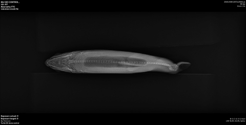
Abernathy Fish Technology Center in Longview, WA, purchased a portable digital x-ray system in fall 2020. The system is composed of a battery-powered x-ray generator and a digital (DR) panel that connects wirelessly to a laptop computer containing image-processing software (see Figure). An x-ray image can be obtained and viewed within a few seconds once the specimen has been placed on the DR panel and exposure has been taken.
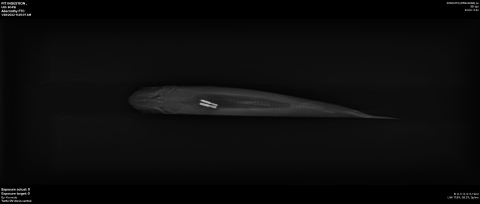
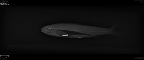
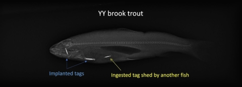
The initial motivation to obtain the x-ray system was to help with a laboratory study on retention of passive integrated transponder (PIT) tag retention in brook trout. We took x-ray images of every fish in the study to confirm tag retention, because sometimes a tag can fail and not be detected by a hand-held reader. Even more importantly (as we found out in real time) x-rays revealed instances where a tag was lost by one fish and eaten by another. We did not expect this, and potentially would have missed this phenomenon if we were relying on just the tag reader, as two of the same kind of PIT tags in close proximity can cause a “tag collision” such that neither tag is detected!
We have another study going, this time with two species of Pacific salmon, to better understand what happens when a juvenile ingests a tag and how long it remains within a fish’s digestive system. Time series x-ray images of fish who have ingested PIT tags is an integral part of this study.
Recently, one of the white sturgeon held at Abernathy became sick and died. As part of the post-mortem, a veterinarian from the Pacific Region’s Fish Health program took x-rays of the sturgeon to see if the images could help diagnose the cause of the fish’s demise. We anticipate that additional, and potentially novel uses of the x-ray system may present itself in the future, and hope that the system can be a resource for other biologists in the Region.